

Introduction
Erythroplakia is a rare but serious oral lesion characterized by a red, velvety patch on the mucous membrane of the mouth. Unlike leukoplakia, which presents as white patches, it is often associated with a high risk of malignant transformation. Due to its potential to develop into oral cancer, early detection and prompt treatment are crucial.
Causes and Risk Factors
The exact cause is not fully understood, but several risk factors have been identified:
- Tobacco Use – Both smoking and smokeless tobacco increase the risk of developing erythroplakia.
- Alcohol Consumption – Chronic alcohol intake can contribute to the formation of oral lesions.
- Poor Oral Hygiene – Inadequate oral care may lead to chronic irritation and mucosal changes.
- Human Papillomavirus (HPV) Infection – Some strains of HPV have been linked to oral cancers and precancerous lesions.
- Nutritional Deficiencies – Deficiencies in vitamins A, C, and iron may predispose individuals to oral mucosal changes.
- Chronic Irritation – Ill-fitting dentures, sharp teeth, or repeated trauma to the oral mucosa can contribute to lesion formation.
Symptoms of Erythroplakia
It may present with the following symptoms:
- Red Patches– A well-demarcated, velvety red lesion that does not rub off.
- Painless Progression– Initially, erythroplakia is asymptomatic, making early diagnosis challenging.
- Ulceration– In some cases, the lesion may develop ulcerations or erosions.
- Burning Sensation– Some patients may experience mild discomfort or burning, especially with spicy foods.
- Irregular Borders– The lesion may appear smooth or slightly raised with irregular margins.
Diagnosis
Erythroplakia is primarily diagnosed through a combination of clinical examination and biopsy. The diagnostic process typically includes:
- Clinical Examination– A dentist or oral healthcare professional inspects the lesion’s appearance, location, and characteristics.
- Biopsy– A tissue sample is taken and examined histologically to determine the presence of dysplasia or malignant transformation.
- Toluidine Blue Staining– A diagnostic aid used to identify high-risk areas that require biopsy.
- Imaging Studies– In cases where malignancy is suspected, additional imaging such as MRI or CT scans may be required.
Treatment Options
The treatment depends on its histopathological findings and severity. Common approaches include:
- Surgical Excision– The preferred method for confirmed dysplasia or carcinoma to prevent malignant transformation.
- Laser Ablation– Used for superficial lesions with no signs of severe dysplasia.
- Cryotherapy– Freezing the affected tissue with liquid nitrogen in some cases.
- Regular Monitoring– Patients with mild dysplasia may require close follow-up to detect any progression.
- Elimination of Risk Factors– Cessation of tobacco and alcohol use significantly reduces recurrence risk.
Prevention and Oral Care Tips
Since it has a high potential for malignant transformation, preventive measures are crucial:
- Quit Smoking and Alcohol– Avoid tobacco and limit alcohol intake to reduce oral cancer risk.
- Maintain Good Oral Hygiene– Regular brushing, flossing, and professional cleanings help keep the oral cavity healthy.
- Routine Dental Check-Ups– Early detection through regular oral screenings can prevent severe complications.
- Balanced Diet– A nutrient-rich diet with sufficient vitamins and minerals supports oral health.
- Address Dental Issues– Properly fitted dentures and treatment of chronic irritation can prevent lesion development.
FAQs
1. What is erythroplakia?
It is a rare but serious oral lesion that appears as a red patch on the mucous membrane. It has a high risk of malignant transformation into oral cancer, making early detection crucial.
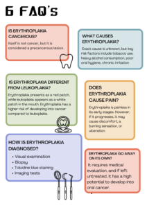
2. Is erythroplakia cancerous?
It itself is not cancer, but it is considered a precancerous lesion. Studies suggest that a significant percentage of erythroplakia cases show dysplasia (abnormal cell changes) or even early-stage cancer upon biopsy.
3. What causes erythroplakia?
The exact cause is unknown, but key risk factors include tobacco use, heavy alcohol consumption, poor oral hygiene, chronic irritation (ill-fitting dentures, sharp teeth), HPV infection, and nutritional deficiencies.
4. How is erythroplakia different from leukoplakia?
It presents as a red patch, while leukoplakia appears as a white patch in the mouth. Erythroplakia has a higher risk of developing into cancer compared to leukoplakia.
5. What does erythroplakia look like?
It typically appears as a well-demarcated, velvety red lesion on the tongue, floor of the mouth, soft palate, or gums. It does not rub off and may have an irregular shape.
6. Does erythroplakia cause pain?
In most cases, it is painless in its early stages. However, if it progresses, it may cause discomfort, a burning sensation, or ulceration.
7. How is erythroplakia diagnosed?
A dentist or oral specialist diagnoses through:
- Visual examination
- Biopsy(tissue sample analysis)
- Toluidine blue staining(to highlight abnormal cells)
- Imaging tests(MRI, CT scan for suspected cancerous lesions)
8. Can erythroplakia go away on its own?
No, erythroplakia does not resolve on its own. It requires medical evaluation, and if left untreated, it has a high potential to develop into oral cancer.
9. How can erythroplakia be prevented?
To lower the risk of erythroplakia:
- Avoid tobacco and alcohol
- Maintain good oral hygiene
- Eat a nutrient-rich diet
- Get regular dental check-ups
- Treat chronic oral irritations (ill-fitting dentures, sharp teeth, etc.)
10. When should I see a healthcare provider?
If you notice any persistent red patches in your mouth that last more than two weeks, especially if they bleed easily, consult a dentist or healthcare provider promptly for evaluation.
Conclusion
Erythroplakia is a potentially serious oral condition with a high risk of malignant transformation. While it may not initially cause symptoms, timely diagnosis and intervention are vital. By understanding its risk factors, maintaining good oral hygiene, and seeking professional care, individuals can reduce their chances of developing this condition. If you notice any persistent red patches in your mouth, consult a dentist or oral specialist immediately to ensure early detection and treatment.
1 thought on “Erythroplakia: Causes, Symptoms, Diagnosis, and Treatment”